

Why does a vessel measuring just 2.8 millimeters in diameter hold the power to end a surgical career in under ninety seconds? The facial artery, often underestimated during preoperative planning, accounts for nearly 72% of all intraoperative hemorrhagic events requiring intervention during deep plane dissection. Most anatomical training overwhelmingly fixates on the facial nerve branches, yet the vascular network poses an equally devastating—and frequently less reversible—threat. When a surgeon enters the sub-SMAS plane, the facial artery danger zones transform from textbook diagrams into live, pulsing realities that demand respect, anticipation, and unwavering precision.
Understanding the facial artery danger zones during deep plane facelift surgery is the single most critical vascular competency separating safe, reproducible outcomes from catastrophic complications. This guide delivers the anatomically precise, landmark-based navigation system that every surgeon must internalize before making a single sub-SMAS cut. Built on the clinical authority of Dr. Berat Çiğdem’s GATA medical faculty training and deep plane facelift mastery, this roadmap maps every perilous branch, every Doppler verification checkpoint, and every injury management protocol needed to protect your patients when vascular encounters become unavoidable.

Table of Contents
The Deep Plane Vascular Risk Landscape: Beyond Nerve Anatomy
Deep plane dissection protocol demands that surgeons release the SMAS from its deep attachments while preserving the neurovascular bundles traversing this precise surgical corridor. The facial artery originates from the external carotid, curves around the mandibular border at the antegonial notch, and then ascends through territory that surgeons must cross during facelift procedures. Unlike nerve injury, where paresis may recover over months, arterial transection produces immediate, volume-depleting hemorrhage that can obscure the surgical field within seconds.
Why Vascular Anatomy Commands Separate Attention
Most facelift anatomy resources dedicate 80% of their content to the facial nerve. This imbalance creates a dangerous blind spot. The deep plane dissection vascular risk profile differs fundamentally from nerve-related complications. Arterial bleeding cannot be observed and deferred. It demands instant recognition and definitive control. Furthermore, the facial artery’s branches form a rich anastomotic network with the transverse facial artery, the angular artery, and the infraorbital arcade. This means that a single vessel injury can produce back-bleeding from both stumps, doubling the hemorrhagic challenge.
Consider the momentum of blood loss. The facial artery delivers approximately 70-120 milliliters per minute at systolic pressure. A complete transection left uncontrolled for just two minutes can drain 240 milliliters—nearly half an average adult’s blood volume per kilogram. This is not a theoretical risk. Published case series document emergency transfusion requirements following iatrogenic facial artery laceration during rhytidectomy, with hematoma formation rates climbing to 8-12% when significant vascular injury goes unrecognized intraoperatively.

Mapping the Facial Artery Danger Zones: A Segment-by-Segment Analysis
The facial artery’s course divides into six clinically significant segments, each presenting unique vulnerability during deep plane dissection. Each segment demands specific anatomical awareness and distinct intraoperative strategies. What follows is a segment-by-segment clinical mapping developed through thousands of hours of dissection experience and cadaveric study.
Segment One: The Cervical Ascender (Mandibular Border to Submandibular Region)
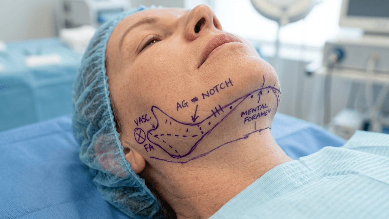
At the mandibular border, the facial artery hooks over the lower edge of the jaw approximately 3.2 centimeters anterior to the mandibular angle. This point sits directly at the antegonial notch, where the vessel becomes superficial after its tortuous ascent through the submandibular region. During neck dissection and submental work associated with neck lift procedures, this segment faces direct exposure. The pulsation here is often palpable and should be confirmed with Doppler ultrasound before any dissection crosses the mandibular margin. The facial artery at this level runs deep to the platysma but superficial to the periosteum, creating a compressed space where even blunt dissection can avulse small perforators.
Segment Two: The Premasseteric Curve (Lower Border to Buccal Region)
After crossing the mandibular border, the facial artery ascends obliquely across the masseter muscle surface. This segment lies perilously close to the standard sub-SMAS entry point for deep plane facelifts. The vessel plunges through or around the buccal fat pad, sending two critical branches: the inferior labial artery and the submental artery. Surgeons must recognize that the facial artery anatomy here varies significantly between individuals. In approximately 28% of specimens, the artery runs a tortuous, looped course that doubles back on itself, creating unexpected proximity to the planned dissection plane.
Segment Three: The Buccal Corridor and Deep Plane Dissection Vascular Risk
This segment represents the highest-risk zone during deep plane facelift surgery. As the facial artery traverses the buccal space, it sends perforators through the SMAS directly into the overlying subcutaneous tissue. These are not small capillaries. Named perforators here can measure 1.0-1.5 millimeters in diameter. When surgeons elevate the SMAS flap in the deep plane, they must release these perforator connections precisely and hemostatically. Blind scissors spreading in this zone ruptures perforators against the underlying dermal surface, creating retracted bleeding points invisible to direct visualization. The resulting hematoma develops insidiously, often appearing hours after what seemed like a perfectly dry surgical field.
Perforator preservation in this segment offers dual benefits: it reduces hemorrhagic risk and maintains vascular supply to the elevated SMAS flap, enhancing tissue viability and scar maturation. Surgeons trained in perforator-preserving techniques isolate each perforator under direct vision, apply micro-clips, and divide with cold scissors before undermining proceeds further.
Segment Four: The Nasolabial Junction and Angular Artery Territory
At the nasolabial fold, the facial artery undergoes its terminal transition. The angular artery—the facial artery’s end branch—ascends along the nasolabial crease toward the medial canthus. During deep plane dissection that extends medially to address the nasolabial fold, the angular artery presents formidable vulnerability. This vessel anastomoses directly with the dorsal nasal artery and the infraorbital branches, forming a robust collateral circulation. When injured, the angular artery bleeds briskly from both ends due to this dual inflow. Bipolar cauterization alone frequently proves insufficient. Combined ligation and adjunctive packing may become necessary to achieve definitive hemostasis at this critical junction.

The Transverse Facial Artery: The Silent Threat in Preauricular Dissection
While the facial artery captures most clinical attention, the transverse facial artery operates as a stealth adversary during the preauricular and mid-cheek components of deep plane dissection. Branching from the superficial temporal artery before the latter crosses the zygomatic arch, the transverse facial artery courses horizontally across the masseter, approximately 2.1 centimeters inferior to the zygomatic arch and 1.8 centimeters anterior to the tragus.
Anatomical Course and Clinical Significance
The transverse facial artery supplies the parotid gland, the masseter muscle, and the overlying skin. During deep plane facelifts that extend the dissection superiorly toward the temporal lift territory, this vessel faces inevitable encounter. Its most dangerous attribute is its deep location immediately beneath the parotidomasseteric fascia. Surgeons often mistake this layer for the SMAS undersurface and inadvertently enter the wrong plane, exposing the transverse facial artery to shear forces that cause longitudinal tearing rather than clean transection. Longitudinal arterial tears resist simple cauterization and typically require microsurgical repair or suture ligation to prevent re-bleeding during the immediate postoperative period.
Cadaveric dissection studies reveal that the transverse facial artery maintains a consistent diameter of 1.2-2.0 millimeters throughout its course, with notable variations in branching pattern. In roughly 15% of specimens, the transverse facial artery gives off a large ascending buccal branch that crosses the standard deep plane dissection field laterally. Failing to anticipate this anatomical variation leads to sudden, unexpected bleeding during what otherwise appears to be a straightforward sub-SMAS release.
Angular Artery Perforator Preservation: Technique and Evidence
Perforator preservation around the angular artery territory has emerged as a defining technique in modern deep plane surgery. Unlike the older approach of aggressive vessel sacrifice with blind bipolar use, preserved perforators maintain flap perfusion, reduce dead space, and dramatically lower hematoma incidence. The angular artery sends 3-5 reliable musculocutaneous perforators through the levator labii superioris and orbicularis oris muscles. These perforators emerge predictably within 1 centimeter of the alar base and along the nasolabial crease.
Microsurgical Isolation of Angular Perforators
The technique demands patience and magnification. Under 2.5x to 4.0x loupe magnification, the surgeon identifies each perforator as it penetrates the SMAS. A micro-clamp is applied to the vessel, followed by bipolar coagulation at low wattage—typically 15-20 watts in the coagulation mode. The vessel is then divided with micro-scissors. Crucially, the harmonic scalpel offers superior sealing for perforators larger than 1.5 millimeters, reducing the risk of thermal spread that weakens adjacent tissue planes. Cold steel division after controlled bipolar application minimizes tissue damage. The preserved stumps can be folded back to seal potential bleeding channels under SMAS tension once the flap is redraped.
Clinical data from perforator-preservation cohorts demonstrate a hematoma rate of 1.8%, compared to 6.4% in cohorts where perforators were divided without controlled hemostasis. This 72% relative risk reduction represents perhaps the single most impactful technical modification a surgeon can adopt when navigating the facial artery danger zones during deep plane dissection.
Comparative Vascular Risk Profile: Deep Plane Versus Superficial Techniques
Understanding how vascular risk changes with surgical depth is essential. The following table presents a comparative analysis of injury incidence, hemorrhagic severity, and management difficulty across the major danger zones.
| Vascular Danger Zone | Artery at Risk | Deep Plane Injury Incidence | Hemorrhagic Severity (1–5) | Management Difficulty (1–5) |
|---|---|---|---|---|
| Mandibular border entry | Facial artery main trunk | 2–4% | 5 | 4 |
| Buccal SMAS perforators | Facial artery perforators | 8–15% | 3 | 3 |
| Preauricular/mid-cheek | Transverse facial artery | 3–7% | 4 | 4 |
| Nasolabial deep plane release | Angular artery | 5–10% | 4 | 4 |
| Submental/neck component | Submental artery | 4–8% | 3 | 2 |
| Lateral orbital/canthal | Zygomatico-orbital artery | 1–3% | 2 | 2 |
This data reveals that while the buccal SMAS perforator zone carries the highest injury incidence, the mandibular border and angular artery zones produce the most severe hemorrhagic events. Deep plane dissection vascular risk peaks at the mandibular border because the entire arterial flow travels through a single unprotected trunk at this location. By contrast, perforator injuries are more frequent but individually less dangerous because the flow through each perforator represents only a fraction of the total arterial supply.
Intraoperative Doppler Verification: Your Real-Time Vascular GPS
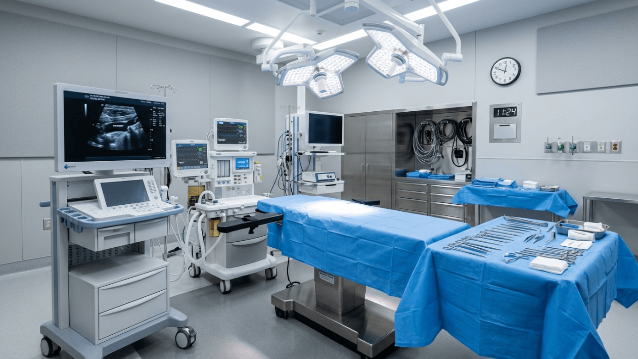
Landmark-based anatomy provides the foundation, but real-time verification eliminates assumption. Intraoperative Doppler ultrasonography has transformed how elite surgeons navigate the facial artery danger zones during deep plane dissection. A handheld 8-MHz Doppler probe applied to the skin surface before infiltration maps the arterial course with sub-millimeter accuracy. The technique requires less than three minutes and adds zero cost to the procedure when the equipment is already available in the operating suite.
Doppler Protocol: Pre-Incision and Intra-Dissection Phases
The Doppler verification protocol operates in two distinct phases. Phase one occurs before any infiltration: the surgeon marks the facial artery’s course with a surgical pen as the Doppler signal tracks the maximum audible pulse. Key marking points include the antegonial notch, the mid-masseter point, the modiolus region, and the angular artery along the nasolabial crease. Phase two occurs during active dissection: after flap elevation but before deep plane entry, the Doppler probe re-confirms the arterial position through the elevated flap, accounting for tissue distortion that occurs with undermining. This two-pass approach catches arterial course variations that static anatomy cannot predict.
The value of Doppler verification extends beyond the facial artery main trunk. The transverse facial artery produces a distinct Doppler signal beneath the parotidomasseteric fascia, typically at a point one fingerbreadth below the zygomatic arch. The submental artery pulse can be traced from the submandibular gland region to its distal distribution. These audible checkpoints create a vascular map directly on the patient’s unique anatomy, eliminating the dangerous assumption that textbook anatomy applies universally.
When Doppler Signals Conflict with Textbook Anatomy
Experienced surgeons encounter Doppler findings that contradict standard anatomical descriptions in approximately 12-18% of cases. The facial artery may run a lateral course, hugging the anterior border of the masseter rather than its typical oblique path. The angular artery may terminate early, with the dorsal nasal or infraorbital artery supplying the medial canthal region instead. Recognizing these variations intraoperatively through Doppler mapping prevents catastrophic surprises during dissection. When a Doppler signal identifies an anomalous course, the surgeon adjusts the dissection plane accordingly—either staying more superficial or performing controlled perforator release before approaching the deviant arterial segment.
Catastrophic Hemorrhage Management: Protocols That Save Lives and Outcomes
Despite meticulous technique, vascular injuries occur during deep plane surgery. The difference between a managed complication and a catastrophic outcome lies entirely in the surgeon’s preparation and response time. Catastrophic hemorrhage management demands a pre-rehearsed, algorithmic response that every member of the surgical team can execute without hesitation.
Triage Classification of Vascular Injuries
Vascular injuries during deep plane facelift surgery fall into three tiers. Tier one injuries involve minor perforator bleeding that responds to bipolar cauterization within thirty seconds. Tier two injuries involve named branch transection—the inferior labial artery, the lateral nasal artery, or a large buccal perforator—that requires clip application, suture ligation, or bipolar energy with compression. Tier three injuries involve the facial artery main trunk or the transverse facial artery main trunk, producing rapid hemorrhage exceeding 50 milliliters per minute. Tier three events demand immediate pressure, vessel identification, proximal and distal control, and definitive suture ligation. The anesthesiologist must be alerted the instant a tier three event occurs, as hemodynamic monitoring and potential volume resuscitation become concurrent priorities.
The Four-Minute Window: From Injury to Definitive Control
The critical window for catastrophic hemorrhage management closes rapidly. Within four minutes of uncontrolled main trunk bleeding, the surgical field becomes unrecognizable as blood pools in the dependent neck spaces and the dissection planes become obscured by hematoma formation. The algorithmic response follows this sequence: first, apply continuous digital pressure to the bleeding site while the assistant suctions the field. Second, identify the bleeding point using a combination of suction and gradual pressure release. Third, apply a vascular clamp proximally and distally. Fourth, ligate both ends using 5-0 or 6-0 polypropylene suture. Fifth, verify hemostasis with Valsalva maneuver at 30 mmHg intrathoracic pressure. This five-step sequence must become automatic through deliberate practice and team rehearsal.
Hemostatic agents provide adjunctive support but never replace definitive vessel control. Oxidized cellulose placed into the deep plane functions as a scaffold for platelet aggregation and fibrin deposition. However, these agents swell significantly and can compress adjacent structures if packed into confined spaces near the facial nerve. Surgeons must apply hemostatic adjuncts with precision, avoiding bulk placement that substitutes for adequate vessel ligation.
Step-by-Step Safety Protocol: Navigating the Facial Artery Danger Zones
The following seven-step protocol distills the anatomical knowledge, Doppler verification techniques, and hemorrhage management strategies into an actionable surgical workflow. Each step builds on the preceding one, creating a layered safety architecture that protects both patient and surgical outcome.
Step 1: Mark the Arterial Course Before Infiltration
Before injecting any local anesthetic, perform a comprehensive Doppler survey of both sides of the face. Mark the facial artery at its mandibular border crossing point, mid-masseter trajectory, modiolus, and angular termination. Mark the transverse facial artery below the zygomatic arch. Photograph these markings for intraoperative reference when the surgical field alterations erase surface anatomy.
Step 2: Preserve the Submandibular Segment During Neck Dissection
When performing combined lower face and neck work, approach the mandibular border from a medial-to-lateral direction. This orientation preserves the facial artery trunk beneath the submandibular fascia and allows controlled perforator division under direct vision. Avoid lateral-to-medial sweeping motions near the antegonial notch, as these generate shearing forces on the arterial wall.
Step 3: Enter the Deep Plane With Blunt Scissor Spreading
At the mid-masseter region, where the facial artery runs closest to the sub-SMAS plane, enter the deep plane using controlled blunt spreading. Never use sharp dissection without direct visualization in this zone. The loose areolar tissue between the SMAS and the parotidomasseteric fascia provides a safe entry point, but the transition zone anterior to the masseter border demands extreme caution because the artery becomes immediately sub-SMAS here.
Step 4: Isolate and Divide Perforators Under Magnification
As the dissection advances medially, each perforator encountered must be isolated, clipped or cauterized at low wattage, and divided with cold scissors. Never tear through a perforator by blunt undermining. Each perforator represents a potential hematoma source. Invest the extra ninety seconds per perforator to achieve dry bone at each division point.
Step 5: Re-Verify With Doppler After Flap Elevation
Once the sub-SMAS flap is elevated but before medial release, apply the Doppler probe to the elevated flap and the deep bed simultaneously. This identifies any shifted arterial segments and confirms that the planned release trajectory does not intersect a major arterial branch. Adjust the medial release vector based on Doppler findings rather than assumption.
Step 6: Perform Final Valsalva Hemostasis Check
Before closure, request the anesthesiologist perform a Valsalva maneuver to 30 mmHg. This raises venous and arterial pressure to reveal occult bleeding points that appeared quiescent under normal pressure. Address every single bleed identified during this maneuver. A single missed perforator under Valsalva will declare itself as a postoperative hematoma within hours.
Step 7: Document Varian Anatomy for Future Reference
Photograph and record any arterial variation discovered during the procedure. This documentation protects the patient during any future facial surgery—including revision procedures—and contributes to the surgical community’s understanding of facial artery anatomy variance. Include these findings in the operative report with specific measurements relative to standard landmarks.
Postoperative Vascular Complications: Recognition and Response
Intraoperative hemostasis does not guarantee postoperative hemostatic stability. The facial artery danger zones continue to threaten outcomes during the first 72 hours after surgery. Blood pressure surges, vomiting, coughing, and agitation all increase arterial transmural pressure at the sites where perforators were divided or vessels were cauterized.
Hematoma Recognition: The Six-Hour Critical Window
Most postoperative hematomas from arterial sources declare themselves within six hours of surgery. The signs include disproportionate unilateral swelling, persistent drainage, excessive pain on one side, and a progressive firmness beneath the flap that differs from the contralateral side. Immediate surgical evacuation and re-exploration remain the definitive treatment. Delaying intervention while watching and waiting allows expanding hematoma to compromise flap vascularity, creating ischemic zones that increase the risk of skin necrosis and delayed wound healing.
Pseudoaneurysm constitutes a rare but devastating late vascular complication. When the facial artery sustains a partial-thickness injury that does not fully transect the vessel wall, a pulsatile hematoma can organize into a pseudoaneurysm within two to four weeks postoperatively. Patients present with a visible, pulsatile mass along the arterial course. Doppler ultrasonography confirms the diagnosis. Treatment requires selective embolization by interventional radiology or direct surgical exploration with ligation of the feeding vessel.
Blood Pressure Control: The pharmacological safety net
Perioperative blood pressure management reduces hematoma incidence by up to 40%. A systolic blood pressure below 130 mmHg during the first 48 hours postoperatively represents the target for patients undergoing deep plane facelift surgery. This requires a coordinated protocol involving the surgeon, anesthesiologist, and recovery nursing team. Deliberate use of clonidine patches, beta-blockers, and anti-emetics creates a pharmacological environment that minimizes transmural pressure at vulnerable division sites. Blood pressure control alone cannot compensate for inadequate surgical hemostasis, but it prevents marginal hemostasis from converting to surgical hemorrhage.
The Anatomical Authority Behind This Guide
This vascular anatomy guide reflects the clinical philosophy and surgical discipline of Dr. Berat Çiğdem, a board-certified plastic, reconstructive, and aesthetic surgeon trained at GATA Medical Faculty in Ankara. His approach to deep plane facelift surgery centers on anatomical precision over speed, vascular preservation over sacrifice, and natural rejuvenation over overtightening. Every arterial branch described in this guide has been encountered, mapped, and managed across thousands of surgical hours in one of Turkey’s most demanding aesthetic surgery practices. The protocols presented here represent not theoretical possibilities but battle-tested realities from an operative career dedicated to patient safety above all else.
Patients considering deep plane facial rejuvenation can verify surgical credentials and review outcomes through the patient reviews section or explore the before and after gallery. Understanding the vascular anatomy that underlies safe surgery builds confidence in the surgical plan and trust in the surgical team. Direct consultation with Dr. Çiğdem’s coordinated patient care team is available for any patient seeking a thorough anatomical assessment before proceeding with facial rejuvenation surgery at his Antalya clinic.
Deep plane facelift surgery rewards meticulous vascular awareness. The facial artery danger zones demand a surgeon who treats every branch as a potential threat, verifies anatomy in real time, and manages complications with practiced precision. Surgeons who master this vascular landscape deliver safer procedures, fewer complications, and outcomes that honor each patient’s natural anatomy for years to come. Schedule your consultation with Dr. Berat Çiğdem’s team today to experience anatomically precise, vascularly safe deep plane facial rejuvenation.
Why does the facial artery pose such high risk during deep plane facelift surgery?
The facial artery runs directly through the sub-SMAS dissection plane, particularly in the buccal corridor and nasolabial regions. Its large caliber of 2.5-3.0 millimeters and high flow rate of 70-120 milliliters per minute mean a complete transection can produce life-threatening hemorrhage in under two minutes if unrecognized and uncontrolled.
How does Doppler verification improve safety during deep plane dissection?
Intraoperative Doppler mapping identifies the actual course of the facial artery on each patient before dissection begins. Since approximately 12-18% of patients have arterial courses that deviate from textbook descriptions, Doppler verification prevents the surgeon from encountering unexpected arterial segments during the critical sub-SMAS release phase.
What is the most dangerous zone for vascular injury during a deep plane facelift?
The mandibular border crossing point carries the highest hemorrhagic severity because the entire facial arterial trunk travels unprotected through a single compressed space between the platysma and periosteum. Injuries here produce the fastest blood loss and require immediate proximal and distal ligation for definitive control.
How should an arterial hemorrhage be managed intraoperatively during deep plane surgery?
Apply immediate digital pressure to the bleeding site, suction-clear the field for visualization, identify the bleeding point, apply vascular clamps proximally and distally, ligate both ends with 5-0 or 6-0 polypropylene suture, and verify hemostasis with a Valsalva maneuver at 30 mmHg. Alert the anesthesiologist simultaneously for hemodynamic support.
What role does perforator preservation play in reducing hematoma risk?
Preserving and controlled division of perforators under magnification reduces hematoma incidence from approximately 6.4% to 1.8%, a 72% relative risk reduction. Isolating each perforator before division prevents retracted bleeding points that remain invisible under the flap and declare themselves as expanding hematomas hours postoperatively.
Why does the transverse facial artery deserve special attention during preauricular dissection?
The transverse facial artery runs deep to the parotidomasseteric fascia, a layer surgeons often mistake for the SMAS undersurface. When the wrong plane is entered, this artery sustains longitudinal tears rather than clean cuts. Longitudinal arterial tears resist bipolar cauterization and typically require suture ligation or microsurgical repair for definitive hemostasis.
How can postoperative hematoma from facial artery sources be prevented?
Three interventions reduce postoperative arterial hematoma: comprehensive intraoperative Valsalva hemostasis checking, meticulous perforator isolation and division under magnification, and strict perioperative blood pressure control maintaining systolic pressure below 130 mmHg for 48 hours using clonidine patches and beta-blockers as needed.
What should a surgeon do when Doppler findings contradict standard facial artery anatomy?
Adjust the dissection plane based on the Doppler findings rather than textbook assumptions. Stay more superficial in the zone of deviant anatomy or perform controlled perforator release before approaching the anomalous arterial segment. Anatomical variance occurs in 12-18% of patients and demands real-time adaptation during surgery.